That Infect Goats
The following information is for informational purposes only. If your goat is ill you should seek veterinary advice. Different species of parasites and their eggs and larvae are difficult for an untrained eye to distinguish from one another. Only a pathologist can accurately identify uncommon or unusual parasites. An understanding of the life cycles of many of these parasites may aid the goat owner in management practices that will prevent outbreaks of parasitic diseases.
Coccidiosis
Eimeria intricata and E. ovina (E. arloingi)
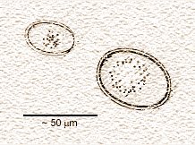 Coccidiosis is a disease that results from the invasion and destruction of the mucous lining of the small intestine by 10 to 12 different species of one celled organisms (protozoa) of the genus Eimeria and one species of the genus Cryptosporidium of the taxonomic Order Coccidiomorpha. The image shows the oocysts of two different species of Eimeria
Eimeria infections can result in serious clinical signs of fluid diarrhea, which may or may not contain mucous or blood, dehydration, emaciation, weakness, loss of appetite, and death. Some goats may instead be constipated and suddenly die without diarrhea. The small oocysts of Eimeria can be found in the thousands in fecal flotation samples. However, diarrhea can appear 1 to 2 days before the eggs and can continue after the oocyst discharge has returned to low levels.
Life History
The life cycle of Eimeria species is somewhat complicated. The oocyts discharged with the feces are uninfective at first. Under the right conditions of moisture and temperature the protoplasm of the oocysts organizes into 4 secondary cysts (sporocysts) which each contain 2 sporozoites. It takes about 2 days for the infective sporulated oocyst to develop. When ingested by the goat the sporozoites escape from the oocyst and invade the mucous lining and cells of the intestine. Once inside the cell, the sporozoite developes into a mass of nuclei which each develop into more sporozoites which enter new cells to repeat the process. It is this constant invasion and destruction of intestinal cells that cause the symptoms of the disease. At some stage, some of the sporozoites form sexual cells which, once fertilized, become the oocytes that are released in the feces. See an animation of the life cycle (requires the Flash plug-in).
Cryptosporidium cause less severe symptoms than Eimeria. Young animals 1 to 3 weeks of age are most susceptible. Symptoms include weight loss, loss of appetite and diarrhea or tenesmus (frequent, futile attempts to empty the bladder or rectum). The disease is seldom fatal. The eggs of Cryptospordium are immediately infective when shed in the feces. Once ingested, the incubation period is about 4 days. Some species that infect calves can also infect man. The eggs of Cryptosporidium are very tiny and transparent-- only 1 tenth the size of an Eimeria oocyst, so are difficult to see without special staining techniques. The disease is self limiting, meaning that with supportive therapy, usually rehydration, the animal recovers on its own.
The Common Thread Worm
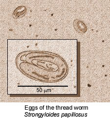
Strongyloides papillosus
The is probably the most common worm that infects goats. More than likely your goat will have a few of these worms most of the time.
Life history
 The adult female worms live in the small intestine where they lay their eggs. The eggs undergo rapid development and are already in the embryonic state when passed out with the feces.
Once outside of the body, the eggs hatch rapidly. After they hatch they can become infective larva or become free-living males and females which can also produce infective larvae. The infectious form completes its life cycle when ingested by a grazing goat or more commonly when the larvae penetrate the skin, usually between the hooves. The resulting skin damage resembles the early stages of foot rot and, in fact may aid in the penetration of the bacteria that cause foot rot.
Once ingested by the goat, the larvae pass into the blood stream and go to the heart, then to the lungs where they emerge into the airways. They work their way or are coughed up to the trachea and finally end up in the intestines where in two to three weeks they develop into mature female worms. The larvae can also be passed from the blood stream into the milk infecting young animals while they nurse. Apparently, the worms are not passed to the fetus in the womb. (see references at the end of this description)
Adults carry a certain amount of immunity to the intestinal form of these parasites, rarely suffering any effects. Sickness or stress however, can cause the worms to take advantage of the situation and build up in large numbers. Heavy infections can cause diarrhea, loss of appetite and weight loss. Continual reinfection results in episodes of coughing as the immature forms migrate to the air passages. If your goat has a large overload, you may see the tiny white worms in the feces.
There is a similar species (Strongyloides stercoralis) that affects humans in many parts of the world. When the larvae penetrate the skin it causes intense itching and the victim may have a brief rise of temperature and a slight headache. As the larvae migrate through the lungs they cause symptoms of lethargy, anorexia, cough and sometimes of mild bronchopneumonia. Humans are not affected by the same parasite that attacks goats but it is interesting to wonder if the goats suffer from some of the same symptoms.
The clear, colorless oval eggs are small and since embryos have usually begun to form by the time the eggs are passed out with the feces they are easily identified. The parasitic females do not produce a large number of eggs so if you see a dozen or so eggs on your slide preparation it represents a large load of worms in your goat.
For more information on this parasite view a few abstracts from scientific papers here.
The Stomach Worms
The large common "twisted" stomach worm
Haemonchus contortus
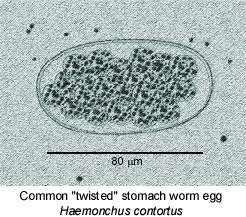 This large stomach worm is 3/4 to 1 1/4 inches long and lives in the fourth stomach (abomasum). The male worm is bright red and the female worm is striped red and white which is why it is sometimes called the "barber pole" worm. The red and white striping results from the white genital organs twisting around the blood-filled intestine.
This parasite is more common in tropical or subtropical areas which has summer rainfall.
Life History
Each female worm is a prolific egg layer, frequently producing 5000 to 10,000 eggs per day. The eggs are already in the early stages of cell division when laid. Once they pass out with the feces the larvae develop to the infective stage in four to five days at the optimal temperature of 75 to 85 degrees F. The pre-infective larvae do not survive long if they dry out but can withstand freezing temperatures for long periods. Once the larvae reach the infective stage they are much more resistant to dry conditions, surviving if not in direct sunlight for weeks. The infective larvae crawl up blades of grass that is wet with dew in the evening then go back down in the morning. In rainy weather they remain on the grass all day, increasing the risk of infection. The goat becomes infected when they swallow grass containing larvae. The larvae undergo several molts in the abomasum to become mature in about 18 days. They will begin laying eggs in three more days.
The large stomach worm pierces the mucous lining of the stomach where they actively suck blood. Severe infestations can cause death of the animal within a week of heavy infection without showing any clinical signs. Animals with chronic infections show anemia and weight loss. The worms tend to infect mostly young animals, however, older animals can develop heavy infections especially during lactation which may prove fatal . In the late stages of infections, the animal develops a swelling beneath the lower jaw, called bottle jaw.
Because the female worm lays such a large number of eggs, the eggs are easily seen using a simple direct smear. Take a small amount of feces, about the size of a match stick tip, smear it on a microscope slide breaking it down with a few drops of water. Place a cover slip over and observe under low power.
A Few More Stomach Worms
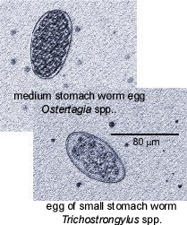
The Medium or Brown Stomach Worm
Ostertagia circumcincta
This worm also lives in the fourth stomach but is much smaller than the twisted stomach worm. Ostertagia circumcincta is only about 1/2 long and is brown in color. More commonly found in temperate climates with winter rainfall and drier summers, the eggs can develop at much lower temperatures often hatching at temperatures lower than 45 degrees F. This makes infections of Ostertagia much more likely during the winter especially during rainfall.
 Life History
The life cycle of the brown stomach worm is similar to the twisted stomach worm. Once the eggs pass out with the feces, the larva hatch and reach the infective stage in five to six days (during the warm summer months). When ingested the larvae make their way to the wall of the abomasum and become coiled in nodules on the mucous membrane. After about a week the larvae leave the nodules and continue to develop into adult worms. Female worms are mature 15 days after initial infection and their eggs are found in the feces just a few days later.
The Small Stomach and Intestinal Worms
Trichostrongylus spp.
Most of the species of the genus Trichostrongylus actually live in the small intestine. Only one T. axei lives in the abomasum. These are very small worms, only 1/4 to 1/3 inch long with thin bodies and are pale pink in color which makes them practically invisible on post-mortem examination. Trichostrongylus, like Ostertagia are more common in areas with winter rainfall.
Life History
When laid, the eggs have already developed to the sixteen or thirty-two cell stage. They develop rapidly, hatching in less than twenty hours in summer temperatures (70 - 80 degrees F.) If the eggs dry out before they hatch they become dormant and can survive for as long as 15 months. In the infective stage, larvae are also very resistant to dry conditions. The goat or sheep is infected when they graze on grass where the larvae have migrated. The larvae travel to the wall of the abomasum or the small intestine where they go through a molt. They then leave the wall to live freely in the lumen of the intestine or stomach. There they develop into adults and are laying eggs within 21 days after infection.
These parasitic worms primarily cause problems in young animals. Adults can carry heavy infections but show little evidence. The intestinal species cause more problems than T. axei which lives in the abomasum. The presence of the intestinal species cause diarrhea, weight loss and loss of appetite. The worms suck blood from the lining of the intestines which causes irritation and swelling of the intestinal membrane. The damaged mucosa results in malabsorption, impaired digestion and protein loss. Heavy infestations may prove to be fatal to the young animal.
The eggs of the various types of stomach worms are very similar. Only a trained eye can distinguish between them. Often, larvae from a dead animal are examined to identify the exact species of the worms.
Nematodirus spathiger
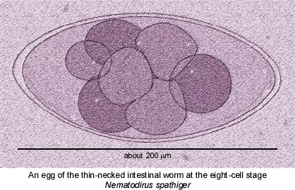
These parasites, so named because the anterior part of the female worm is narrow and coiled, are about 1/2 to 2/3 inch long, and are creamy to bright pink in color. They live in the small intestine.
Life History
When laid, the large eggs are in the early stages of cell division (4 to 8 cells). After passing out with the feces the embryo develops very slowly often taking 14 days in optimum conditions. The larva goes through several molts before they hatch out of the egg. The eggs are very resistant to drying and low temperatures, surviving many months. The goat is infected when they ingest the developing eggs or the large infectious larvae that have crawled up onto grass. The worm matures into an egg-laying adult after about 4 weeks.
Infection is primarily in very young animals, but older animals may also be affected. Because the eggs only hatch in wet conditions, outbreaks usually occur in the spring 2-4 weeks after a rainfall. The infected animal has a sudden onset of diarrhea, dehydration, and general unthriftiness with death occurring 2-3 days later. The eggs are easy to identify, however the animal may show signs of the disease before the female worm has started laying eggs. In addition, the worm lays few eggs, so even with a heavy infestation few eggs may be seen on the preparation.
The Large Lung Worm
Dictyocaulus filaria
These long (up to 4 inches) white, thread-like worms live in the air passages of the lungs. The female worm lays her eggs in the air passages which are coughed up into the pharynx then swallowed. By the time they reach the intestines the first stage larva have already hatched. Some eggs may be coughed out in mucous onto the pastures.
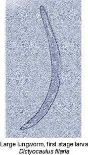
Life History
These larva are stout, with a blunt tail and a round knob at the head end which closes the mouth so that the larvae cannot feed. After one or two days the larvae undergo the first molt, but the skin is not shed and encloses the second-state larva. The larva will undergo another molt in three or four more days again retaining the skin. These larva can survive for up to nine months in shallow water, where they are swallowed by grazing animals. After they are swallowed the larva bore into the wall of the small intestine and travel to the lungs through the lymph vessels. After a third molt, the larvae make another trip through the lymph system to the heart and then back to the lungs through the blood. By then they are fourth stage larva of about 1/50 of an inch long and will be found in the bronchi after eight days. Adults develop in the lungs 18 days after infection and they will produce eggs in 3 to 5 weeks.
Coughing is the most common symptom, beginning about 30 days after infection. Severe infestations can cause pneumonia and suffocation.
Since lungworm eggs hatch before being passed in the feces the eggs generally are not seen by the flotation method. Eggs may be present in the mucous discharge from the nose or be coughed out through the mouth.
To see the larva, float a few fecal pellets on the surface of water in a small glass container. After a minute or two hold the container up to a strong light and the 1 mm sickle-shaped larva can be seen with the naked eye as they swim out and fall through the water. After a few hours draw out some of the water place it on a slide and observe the larvae under low power.